
|
| Malmodelera Program to Interactively Construct a Shape Model from a Three-Dimensional Medical Image |
|
|
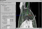
Malmodeler is a program that can be used to interactively construct a shape model from a three-dimensional medical image obtained by computer tomography (CT) or magnetic resonance imaging (MRI). A function of this program enables the superimposition of the shape model, which is being constructed, as a volume image on the medical image. By using a recently available cheap and powerful computer graphics hardware, this program can be operated at a practical speed on the commonly used personal computers. Since window parameters such as brightness, contrast, and threshold are interactively adjustable, and projection operators such as over and maximum intensity projections (MIPs) are also interactively changeable, running blood vessels and shapes of organs can be displayed stereoscopically. Thus, intuitive modeling by utilizing human spatial cognition can be materialized. A multi-resolution mesh, centerline mesh, and tubular mesh that are defined by the shape of the centerline’s orthogonal cross-section can be edited as the objective model based on a polygonal mesh and subdivision surface of arbitrary topology (Fig. 1). Detailed information is available at ( --> Download pages of (1), (2), (3)-(9)).
|
|
|
| Fig. 1 Centerline-based modeling obtained using the multi-resolution mesh in Malmodeler |
|
 |
|
|
CREAM—an Integrated System for a Faster Risk Evaluation of Various Angiopathies
|
|
|
|
|
|
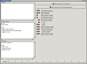
Fig. 2 The tentative user interface of CREAM (the screen is under development)
|
|
|
|
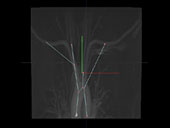
| Fig. 3 Topology of the extracted blood vessels |
|
Although the time required for modeling is shortened by using Malmodeler, 10-60 min is still required for aortic arch modeling. Moreover, environmental conditions required for applying the model in clinical fields have not been established because the calculation time for modeling could not be shortened. In order to avoid modeling and calculation in a clinical setting as far as possible, we aimed to develop CREAMan integrated system for faster risk evaluation of various angiopathies.
The essential requirements for using CREAM are obtained as follows:
| • |
A database to store the calculation models and calculation results is included in the system. Calculation models applicable for various running blood vessels and shapes have been previously constructed and calculated. |
|
| • |
Topology of blood vessel bifurcation or geometric characteristics can be extracted from a blood vessel image of a new patient. Based on the extracted parameters, a similar model and calculation result can be searched in the database and visualized. |
|
| • |
A new shape model can be constructed, and a calculation model can be constructed and calculated, if required. A prototype that was semiautomatically constructed from an image of an individual patient can be used to construct the model. |
|
| • |
A consistent user interface has been prepared on which topology can be extracted from an imported image, and sequential workflow, including model search, model construction, mesh generation, calculation, and visualization of results, based on the topology can be supported. |
|
| • |
Existing modalities such as CT and MRI can be cooperated through digital imaging and communications in medicine (DICOM) |
|
| Currently, a schematic design has been prepared, architectural implementation has been performed, and an implementation test of the user interface has been initiated (Fig. 3). Detailed information is available at ( --> Download pages of (10)-(16)). |
|