
|
| [1] Effect of an elastic body on fluid resistance |
|
|
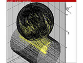
We studied the effect of an elastic cylinder on the fluid resistance to a two-dimensional uniform flow ( --> see download pages (11) and (12)).
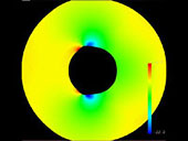
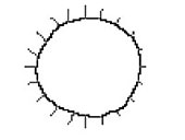
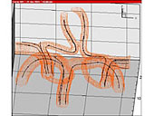
The results obtained were as follows. When the elastic body was soft, traveling waves appeared around the surface of the elastic cylinder (Fig. 1), and the fluid resistance increased. When the elastic body was hard, the fluid resistance decreased in a region just above the critical Reynolds number, although the decrease was small compared to that in the case of a hard body (Fig. 2) <3>. |
|
| [2] Simulation of blood flow in the left ventricle of the heart |
|
|
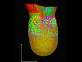
We developed a method to model the left ventricular wall motion and hemodynamics that occur during one heartbeat cycle using data extracted from echocardiographic images of the heart <2>.
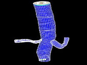
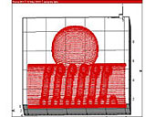
Using this model, we conducted a numerical blood flow analysis based on the finite volume method. A grid was generated using the structured grid method (Fig. 3).
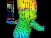
The inlet boundary condition of blood flow velocity was determined from data that was measured using ultrasonic waves. Vortices were found to be generated at the center of the left ventricle (Fig. 4). |
|
| [3] Simulation of blood flow through the renal artery and abdominal aorta |
|
|
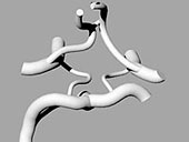
We developed an automatic method for extracting vascular contour data of the renal artery and abdominal aorta from CT medical images of the abdomen <1>.
The method comprises the following two steps: GA (genetic algorithm)-based automatic pattern recognition of the vascular area (Fig. 3) and snake-based extraction of contour data (Fig. 5). Using the extracted data, we constructed a model of the renal artery and that of the abdominal aorta.
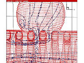
Further, a grid was generated based on the model by using the structured grid method (Fig. 6). A numerical analysis was conducted by using the finite volume method based on a domain decomposition. A swirling flow was found to be generated (Fig. 7). |
|
| [4] Study of voxel model construction by using V-CAD |
|
|
Owing to CAD, models can now be constructed in an interactive manner. Using V-CAD, we developed a rapid method for directly constructing a voxel model from the solid model.
|
| [5] Simulation of extensive blood flow through the major portion of the cerebral aorta |
|
|
We developed a method for constructing a solid model of the cerebral aorta (Fig. 8) by using CT medical images of the cephalic portion ( --> see download page (9)). Subsequently we constructed a voxel model by using V-CAD.
Using a finite-difference scheme in the frame of a rectangular coordinate system, we conducted a numerical blood flow simulation. Our method enabled simulation of the blood flow through the major portion of the cerebral aorta, despite its complex shape (Fig. 9). In addition, we developed a method for extracting the data of the structural line of the cerebral arterial vessel.
Using the multi-block method of the finite volume method, we conducted a numerical blood flow analysis (Fig. 10). |
|
| [6] Simulation of blood flow during intravascular surgery of a cerebral aneurysm by using GDC |
|
|
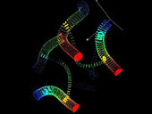
Using CAD, we modeled a catheter, coil, stent, and balloon. On the basis of these instrument models, we constructed a voxel model for each of these by using V-CAD (Fig. 11). Using the finite difference scheme in the frame of a rectangular coordinate system, we conducted a numerical calculation. It was found that the generation of vortices in a cerebral aneurysm can be suppressed by placing a stent. In addition, no vortices were generated by the placement of coils (Fig. 13). Thus, our method was effective for simulating the blood flow during intravascular surgery.
|