
|
Establishment of an Accurate Method to Evaluate Blood Flow Velocity and Vascular Shape Obtained by Magnetic Resonance Imaging (MRI)
—Phase Contrast and Time-of-Flight Methods— |
|
|
The behavior of endothelial cells under conditions of shearing stress and the intramural stress generated in the vascular wall are considered to play an important role in vascular diseases. Therefore, the velocity distribution of blood flow and the vascular shape, which are directly associated with the shearing stress and the intramural stress respectively, are considered essential to understand vascular diseases.
The phase contrast method is the imaging sequence used for noninvasive measurement of blood flow velocity. The signal phase in the method is proportional to the blood flow velocity. Velocity Encoding (VENC) is the velocity corresponding to 180 degrees. The relationship is described as follows: V = VENC * φ/180. (V, velocity; φ, phase). Many studies have been performed to verify the measurement accuracy [1]-[5] The relationship between the velocity and acceleration at the measuring point and the accuracy of the measurement were mainly evaluated in these studies. Their results indicated that the phase dispersion influence the measurement accuracy.
Since the phase contrast method was based on gradient echo method, the signal value is influenced by sequence parameters such as TR and TE, and the relaxation times. However, the influences on the accuracy of velocity measurement have not been adequately evaluated. The typical sequence used for vascular shape evaluation is time-of-flight. In this method, the signal of the flow velocity is higher in the direction perpendicular to a slice (inflow effect). Therefore, the general tendency of the flow velocity can be estimated by using this characteristic. However, this is merely a tendency based on the signal level, and the actual flow velocity can not be measured; thus, this is different from the phase contrast method in which the actual flow velocity is measured.
Therefore, it is considered that in an image obtained by the time-of-flight method, the recognition of the vascular shape in a region can be determined by the impression of the image. However, the evaluation of the shape based on the impression becomes more difficult as the object becomes more complicated. Moreover, it is important to quantitatively evaluate the magnitude of false-positive rate of the region-recognition method by using dynamic simulation based on the shape obtained. It is also considered that if the stationary region in the phase contrast method can be discriminated from the active region by quantitative parameters such as the flow velocity, the magnitude of false-positive rate in the phase contrast method can be quantitatively evaluated. Therefore, the characteristics of the phase in the phase contrast method must be evaluated.
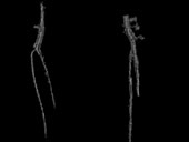
In this study, the influence of the sequence parameters and the relaxation times on the signal phase in the phase contrast method was investigated. The followings were evaluated: (1) Influence on the measured value of the flow velocity (evaluation at the point at which maximum value of the flow velocity, where the influence of the phase dispersion is limited) [6] and [7], (2) influence on the velocity distribution [8] and [9], and (3) signal phase characteristics in the stationary region [7] and [8]. Additionally, the time-of-flight method was applied to the lower extremity, and the tendency of blood flow velocity in the superficial and deep veins was evaluated [10].
The results obtained were as follows: (1) When only the sequence parameters or the relaxation times were altered, there was also a change in the signal phase, (2) the velocity profile was affected by changes in the sequence parameters, (3) the phase characteristics in the stationary region were not included in the average value of the phase but were included in the standard deviation in the region, (4) using the phase characteristics in the stationary region, the popliteal artery could be extracted [7], and (5) using the time-of-flight method, individual differences in the venous blood flow of the lower extremity were observed. Further, changes in the venous blood flow before and after exercise were also noted.
|
|
|
 |

| [1] |
Ku,
D.N., Biancheri, C.N., Pettigrew R.I., Peifer,
J.W., Markou, C.P. and Engels H., Evaluation
of magnetic resonance velocimetry for steady
flow, Transactions of the American Society
of Mechanical Engineers, Journal of Biomechanical
Engineering, 112(1990), 464-472. |
| [2]
|
Oshinski,
J.N., Ku, D.N., Bohning D.E and Pettigrew,
R.I., Effects of acceleration on the accuracy
of MR phase velocity measurements, Journal
of Magnetic Resonance Imaging, 2(1992), 665-670.
|
| [3] |
Siegel, J.M., Oshinski,
J.N., Pettigrew, R.I. and Ku, D.N., Comparison
of phantom and computer-simulated MR images
of flow in a convergent geometry: Implications
for improved two-dimensional MR angiography,
Journal of Magnetic Resonance in Imaging,
5(1995), 677-683. |
| [4]
|
Steinman,
D.A., Ethier, C.R. and Rutt, B.K., Combined
analysis of spatial and velocity displacement
artifacts in phase contrast measurements of
complex flows, Journal of Magnetic Resonance
Imaging, 7 (1997), 339-346. |
| [5] |
Finnie, M., Fullerton,
G.D. and Cameron, I.L., Molecular masking
and unmasking of the paramagnetic effect of
iron on the proton spin-lattice (T1) relaxation
time in blood and blood clots, Magnetic Resonance
Imaging, 4 (1986), 305-310. |
| [6] |
Kato, Y. and Himeno,
R, Extraction Method for Blood Vessels, Based
on the Velocity Profile Measured by Phase
Shift Method,*********??????, (2002-8), 201-208.
|
| [7]
|
****** |
| [8] |
****** |
| [9] |
****** |
| [10] |
Kato, Y. and Himeno,
R, Relationship between the structure and
the velocity profile in the accompanying vein
of the limb,******????????, (2002-8), 209-212. |
| ********************* |
|
|
|