
|
| Blood Flow Analysis Using an Orthogonal Coordinate System |
|
|
Medical image data obtained from devices such as magnetic resonance imaging (MRI), X-ray, and computer tomography (CT) are given as pixels or voxels. Taking advantage of the orthogonal nature of these data, I have been constructing a computational method for conducting a blood flow analysis inside complex structures, using an orthogonal coordinate system. This research was undertaken with the aim of reducing the time required to attach a mesh and of enabling analysis by directly capturing the unique characteristics of individual blood vessels. Owing to this research, computational results for 2-dimensional blood flow analysis were obtained for the blood flow inside a coarctated vessel involving pulsation and inside the carotid artery. The computational results for 2-dimensional analysis inside the coarctated vessel with the pulsating flow and inside the carotid artery are introduced below.
The computational method employed in this study is based on the volume of fluid (VOF) technique developed by Hirt (1981) [1]. In this technique, the coordinates of the boundary points are determined and the unknown variables for the velocity and pressure are assigned to the points on the grid as well as on the boundary points. Further, the study employs the velocity-pressure coupling format, and the technique presented by Nishida (1996) [2] is used in principal for discretization. Nakano’s neighboring point local collocation (NPLC) technique (1995) [3] is used for the points adjacent to the boundary, and the successive over-relaxation (SOR) method is used for the Poisson equation of pressure. In addition, the second-order Adams-Bashforth method is used for the flow velocity term, and the first-order forward Euler method for all the other terms in order to achieve discretization along the time axis. Furthermore, an upwind difference is considered in the advection term, and an original method combining the Neumann condition and the fundamental equations is used for the boundary condition of pressure.
|
|
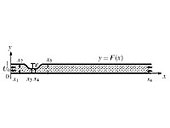
| Fig. 1 Computational stenosis model. |
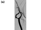
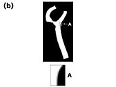
Fig. 2 Data conversion.
(Fig. 2(a): Original image, Fig. 2(b): Enhanced image) |
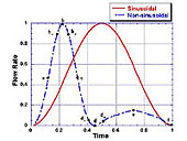
The following are some numerical examples. The first example (Example 1) presents the computational result of the blood flow inside a vessel [4],[5] shown in Figure 1. The second example (Example 2) presents the computational results of the blood flow based on an actual medical image data of the carotid artery (Figures 2(a) and 2(b)). A common assumption of no noise is made for both the examples. For the given shapes, a mesh of width h along the x-axis and height k along the y-axis is used. Each pixel data is applied to the mesh according to the area ratio of the region, and computation is performed based on this pixel data. The influx data shown in Figures 3 [6] and 4 [7] are used for Examples 1 and 2, respectively.
|
|
| Fig. 3 Graph of incoming flows. |
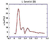
Fig. 4 The flow form in the carotid artery by Olufsen (2000). |
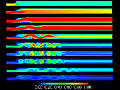
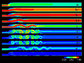
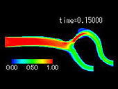
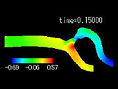
The results for Example 1 are shown in Figures 5 and 6 and those for Example 2 are shown in Figures 7, 8, and 9. With regard to Example 2, the vessel diameter and the maximum influx volume are D = 0.8 cm and Q_p = 30 cm3/s, respectively. The period of pulsation is T 1.1 s; viscosity, μ = 0.049 g/(cm·s); and density, ρ = 1.055 g/cm3[7]. Furthermore, time is represented by the dimensionless variable t.
|
|
| Fig. 5 Isovelocity contours at Re = 750, St = 0.024 with ε= 0.5 in the sinusoidal case. |
Fig. 6 Isovelocity contours at Re = 750, St = 0.024 with ε= 0.5 in the non-sinusoidal case. |
|
|
| Fig. 7 Isovelocity contours at t = 0.15 |
Fig. 8 Pressure contours at t = 0.15 |
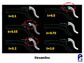
Fig. 9 Streamlines |
|
 |

| [1] |
C.
W. Hirt and B. D. Nichols, Volume of fluid
(VOF) method for the dynamics of free boundaries,
J. Comp. Phys., 39(1981), 201-225. |
| [2]
|
****, 2646-2651. |
| [3] |
****, 4319-4326. |
| [4]
|
H.
Liu and T. Yamaguchi, Effects of Pulsation
and Geometry on Post-Stenotic Oscillatory
Flow, JSME Int. J., Series C, 42, No.3(1999),
612-620. |
| [5] |
H. Liu and T. Yamaguchi,
Waveform Dependence of Pulsatile Flow in a
Stenosed Chnannel, ASME J. Biomech. Ing.,
123(2001), 88-96. |
| [6] |
O. R. Tutty, Pulsatile
flow in a constricted channel, ASME J. Biomech.
Eng., 114(1992), 50-54. |
| [7]
|
M. S. Olufsen, C, S.
Peskin, W. Y. Kim, E. M. Pedersen, A. Nadim
and J. Larsen, Numerical simulation and experimental
validation of blood flow in arteries with
structured-tree outflow conditions, Ann. Biomed.
Eng., Vol.28(2000), 1281-1299. |
|
|