
Lung compliance is one of the important parameters influencing the dynamic phenomena of the airway as well as one of the indicators of respiratory function in clinical medicine. Lung compliance is determined by the elasticity of the lung tissue and the surface tension of the airway. Although lung compliance has been used to evaluate the entire lung macroscopically, respiratory disorders develop in various locations within the bronchioles. Moreover, the airway is not a uniform tissue. In this study, we focused on localized lung compliance and evaluated the bronchiolar compliance. In particular, bronchioles with an inner diameter of 150–300 μm were evaluated. Since the bronchioles were previously enucleated, the results obtained were actually affected by the surrounding lung tissue.
In this study, enucleated rat lungs were used to develop a method of visualizing the bronchioles in the lung parenchymal tissue for evaluating the state of the soft tissue within the bronchioles. Additionally, changes in the morphology and localized compliance during breathing were investigated. |
|
|
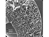
| Fig. 1 Representative micro-CT image showing many small airways and alveoli at FRC. Arrows indicate airways (white: small airways and black: alveoli). Stars indicate blood vessels. Bar: 500 mm. |
|
|
|
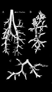
Fig. 2 Three-dimensional reconstruction of the bronchial tree from cross-sectional images using an isosurface approach in VTK.
(A): The entire airway as reconstructed from micro-CT images by using the SCT method (1 cubic voxel size: 43 mm). (B and C): the small airway (1 cubic voxel size: 16 mm, diameter range: 300–170 mm, and Z: 10–16). Small arrow in A indicates the starting point of (B) and (C). |
|
The visualization method developed in this study for the evaluation of the bronchiole involved perfusion of the blood vessel with a contrast medium into. Consequently, the contrast medium leaking from the blood vessels stained the lung tissue. Enucleated lungs were obtained from 14 male Wistar rats (300 ± 30 g) aged 9–10 weeks. A solution of sodium diatrizoate (0.8 g/ml) was used as the contrast medium and perfused into the blood vessel where it remained for 1 h. Subsequently, the lungs were enucleated. A cone-beam-type micro-CT with high spatial resolution was used as the visualization device to observe the bronchioles.
This method enabled the visualization of the bronchioles under conditions that were similar to physiological conditions; thus, a three-dimensional structure of the bronchioles was constructed. Additionally, the morphological characteristics of the bronchiole were quantified. Using a three-dimensional thinning processing, a skeleton of the bronchial tree was extracted, and the length, diameter, and turnout angle of each bronchial generation (Z) were calculated. It was observed that the length and diameter of the bronchioles decreased exponentially against Z. It is already known that asymmetry of branches has a substantial effect on the dynamic phenomena of the trachea. Further, it was observed that the bronchial asymmetry became smaller as Z increased, and the bronchioles branched off more symmetrically as the branching approached closer to the alveolus.
Since this method enabled the visualization of the state of soft tissue, any morphological changes in the same bronchiole could be traced. |
|
|
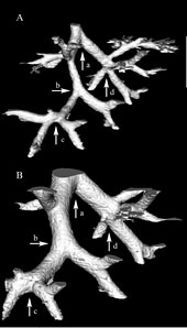
| Fig. 3 Three-dimensional structures of the same branching network at (A) FRC and (B) TLC. Diameter range at FRC: 300–170 mm. Airway generation (Z) range: 10–16. The arrows (a–d) indicated the same dividers in (A) and (B). |
|
|
 |
|
|
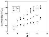
| Fig. 4 Localized compliance at TV and TLC (average ± SE.) as a function of Z. *p (between TLC and TV) < 0.05. |
|
By using a newly proposed method, the morphological changes in the same bronchiole were measured at different lung volumes, and localized lung compliance was evaluated.
The enucleated lungs that were used in this experiment were obtained from 15 male Wistar rats (300 ± 30 g) aged 9–10 weeks. When compared at the time of functional residual capacity (FRC), the rate of increase in bronchiolar length (dL) at the time of tidal volume (TV) and total lung capacity (TLC) were 18% and 43%, respectively. Moreover, the rate of increase in bronchiolar diameter at the time of TV and TLC were 36% and 89%, respectively. In particular, the microscopic diameter–pressure curve showed hysteresis in the case of the bronchioles with a diameter less than 300 μm, and the bronchiolar diameter increased sharply at a certain pressure; this is called a “pop–open” phenomenon. The reason for this is considered to be as follows: (1) at low pressures, the diameter of the bronchioles increases gradually because the balance between the elasticity of the airway wall and the surface tension of surfactant is maintained, and (2) when the pressure becomes greater, this balance is lost and the diameter increases sharply. |
The localized compliance of each generation was measured between the time of FRC and TV (CTV), and between the time of FRC and TLC (CTLC).
Although both CTV and CTLC increased as Z increased, their curves were not linear, that is, the curves of CTV and CTLC were concave and convex, respectively. These results indicate that there is no uniformity in the bronchiolar deformation and that the deformation occurs with phase-shift during breathing.
The localized compliance of the bronchioles clarified in this study can be used as important information for the purpose of gas exchange simulation of the lungs. |
|