
|
|
|
|
Scleral buckling surgery, wherein the eyeball is externally compressed, has been used as a surgical treatment for retinal detachment. However, since the prognosis is largely affected by the extent of eyeball deformation, preoperative quantitative estimation of the deformation is required.
Our study aims to estimate the optimal extent of eyeball deformation by using finite element analysis so that this data can be used for surgery.
The eyeball consists of various soft tissues such as the sclera, choroid, retina, cornea, crystalline lens, and vitreous body. The shape of the eyeball is maintained by the intraocular pressures and the pressure exerted by the surrounding tissues such as the muscles, fat, and nerves. As the dynamic simulation of an eyeball, analysis of collision-induced eyeball injury and that of the cornea and crystalline lens for correcting visual acuity have been mainly studied. Although studies have reported the dynamical characteristics largely related to retinal detachment, there are almost no detailed reports on the dynamical characteristics of tissues such as the sclera, choroid, and retina. In this study, the dynamical characteristics of each eyeball tissue were measured using a tensile test to simulate all the eyeball tissues. |
|
|
|
|
| A swine eyeball was used in this study because obtaining a human eyeball is difficult, and the size of a swine eyeball is similar to that of a human eyeball. The dynamical characteristics of the cornea, sclera, choroid, and lens capsule were measured using a newly developed tensile test system (--> Hideo Yokota’s home page). In this test, we used ring-shaped specimens of each tissue that were sliced with a perforation device (--> a list of related papers (1)). Since the simulation model was assumed to be a hyperelastic body, the measured dynamic data were converted to a nominal stress–strain relationship. Subsequently, the dynamical characteristics were examined for the tissue’s dependence on the environmental temperature, tensile speed, eyeball position, and existence of anisotropy. |
 |
|
|
|
|
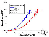
The ring-shaped specimens of each tissue could be obtained using the perforation device. Moreover, the dynamic data under conditions to that of a living body could be obtained using the tensile test performed in a culture solution (-->a list of related papers (1)–(3)). Dependence of the scleral tissue dynamics on the environmental temperature was observed (Fig. 1).
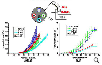
In both the scleral and choroidal tissues, although the deoendence of their dynamical characteristics on the eyeball position was observed, a correlation between these tissues was not recognized (Fig. 2).
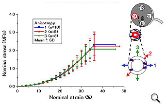
Since collagenous fibers constitute the main scleral component, the existence of anisotropy was examined; consequently, absebce of anisotropy was confirmed (Fig. 3). |
|
|
Fig. 1 Dependence of the scleral tissue dynamics on the environmental temperature
(Click here for detailed information) |
|
|
|
Fig. 2 The dynamical characteristics of the scleral and choroidal tissues with regard to the different positions of the eyeball
(Click here for the detailed information) |
Thus, it was clarified that the dynamical characteristics of each eyeball tissue are different, and that the amount of stress was different from the amount of strain for each tissue when the tissue was ruptured. Since the scleral tissue dynamics were found to be dependent on the environmental temperature, it is considered that acquisition of the dynamical characteristics of each tissue with regard to the environmental temperature, similar to that in a living body and during an operation, is required. Moreover, since the eyeball has a lamellar structure, the tensile test of the adhered complex tissues and an exfoliation test between the adhered tissues are considered necessary to obtain their dynamical characteristics, similar to that in a living body. |
|
|
Fig. 3 Existence of anisotropy in the sclera
(Click here for the detailed information) |
|