
|
| Basic Study of an in vivo Experimental Model for Cortical Bone Remodeling |
|
|
1. Abstract
Although dynamic remodeling of bone tissue is widely regarded as the conventional method to investigate functional adaptation of biological tissues, no biodynamic experimental evidences exist that are sufficiently reliable to elucidate the quantitative relationship between the dynamic load on the bone and its influence on bone remodeling.
In this study, we aimed to develop in vivo experimental models to quantitatively evaluate remodeling of the cortical bone that plays an important role as a structural support of the bone. Therefore, we developed an experimental system wherein neogenetic bone tissue with a known load hysteresis could be induced in vivo. Therefore, we developed flame type devices and implanted them in the tibia of rats for neogenetic bone formation in the devices. Additionally, neonatal bone induction experiment was performed by applying the bone lengthening technique that is widely used in orthopedic treatments.
2. Experimental Devices and Procedures
During the developmental or neogenetic growth, the bone tissue is simultaneously affected by both modeling and remodeling due to dynamic environmental changes. In order to evaluate bone remodeling, it is considered necessary to obtain samples with a known load hysteresis. It is almost impossible to collect the samples under homogeneous conditions while performing in vivo experiments on remodeling using a bone tissue, since the initial conditions of all samples are different.
In this study, we investigated a method to form bone tissue with a known load hysteresis by inducing neogenetic bone tissue in vivo and control the load that affected the process of bone formation.
2.1 The experimental models using flame type devices
In this experiment, to determine the experimental region, the following three experimental models with three types of flames were implanted in the bone and bone tissue was induced into the flames.
(1) Two plates having dimensions of 15 × 2 × 0.5 mm were implanted in the bone parallel to the diaphyseal axis.
(2) Two plates having dimensions of 2 × 2 × 0.5 mm were implanted in the bone parallel to the diaphyseal axis.
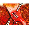
(3) A quadrangular plate with an opening of 10 × 2 mm was implanted in a block having dimensions of 12 × 3 × 2 mm.
We considered the possibility of investigating the influence of the osteon orientation on bone induction by using the experimental models (1) and (2). Additionally, we considered the possibility of investigating bone induction under conditions when the induction region was isolated from the surrounding bone tissues by using the experimental model (3). In order to investigate the basic neogenetic ability of the bone tissue, an experimental model (4) with isolated bone defect was prepared.
The experimental procedure is described below.
Rats (Std: Wistar/ST) were used as the experimental animals. Under anesthetization, flames were implanted in the right tibia. In the experimental model (1), the distance between two plates was set at approximately 1 mm. In the experimental model (2), the distance between two plates was set at approximately 10 mm, and the width of the defect was set at approximately 1 mm. In the experimental model (4), the width of the defect was set at approximately 1 mm. Figure 1 shows the implant operation of the experimental model (3).
Three rats were used for each experimental model. After the operation, rats were bred in a cage and allowed to move freely. |
|
2.2 An external fixation device
In orthopedic practice, a bone lengthening technique using an external fixation device for treating diaplasisbone tissue lost due to tumor enucleationcan be used for bone regeneration; thus, it improve the structural imbalance in the skeleton [1]. In this study, based on the results of experiment [2] performed by Richards et al., an external fixation device for rat was developed and the bone lengthening was performed (the experimental model (5)) using this device.
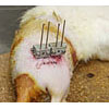
The detailed description of this device is as follows: (a) Essentially, the device consists of two blocks, (b) the two blocks are connected to each other by two guides and a screw, (c) the distance between the blocks can be adjusted by rotating the screw, and (d) the blocks are connected to the bone by pins (Kirschner wires that are 1 mm in diameter).
|
|
The experimental procedure is described below.
Under anesthetization, the pins were inserted into the right tibia of each rat (Std: Wistar/ST); subsequently, the external fixation device was installed. Then, the skin that was approximately 5 mm dorsal to the pin insert position was cut using a knife in the direction parallel to the long axis of femur to expose the femur bone. The femur was cut into two pieces using a micro-surgical saw. In order to achieve contact between the two cut surfaces, the distance between the two blocks of the device was adjusted by using the screw. Then, the severed part was sutured (Fig. 2). After the operation, the rat was bred in a cage and administrated appropriate dosage of antibiotics. After 6 days of breeding without any restriction of movement and food intake, the distance between the two blocks was extended twice a day for 4 days; each time, the gap was extended by a distance of 0.25 mm; thus, the gap was extended by 0.5 mm every day. Thereafter, they were bred again for 4 days without increasing the distance between the two blocks. Subsequently, they were sacrificed to observe the internal structure of the bone using the micro X-ray computed tomography (CT).
|
 |
3. Experimental Results
In the experiments using the models (1)–(3), the neogenetic bone could not be induced in every experimental model because either the flame was detached from the bone, or the bone was fractured near the defective area. In the experiment using the model (4) in which the flame was not implanted, the defect was apparently closed 4 weeks after the operation.
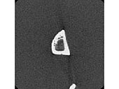
Four and eight weeks after the defect was carved, the rats were sacrificed to observe the internal structure of the bone using the micro X-ray CT. The observation results of 4 and 8 weeks after the induction of the defect are shown in Figs. 3 and 4, respectively.
At the end of 4 weeks, although the tibia surface was apparently induced, holes and cancellous bone-like tissues were observed. Since the experimental region is the central portion of the tibia diaphysis, the presence of cancellous bone tissue is unusual. Therefore, it was considered that this cancellous bone-like tissue was the precursor of the woven bone that would have been formed during the neogenetic process and that there was incomplete regeneration of cortical bone tissue.
At the end of 8 weeks, although the cortical bone regeneration at the defect region was confirmed, the thickness of the regenerated cortical bone was thinner than that of the natural cortical bone in the regions adjacent to the defect region of the right tibia when compared with the corresponding regions in the left tibia. Moreover, the holes and cancellous bone-like tissues were persistent in the defect region. |
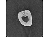
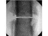
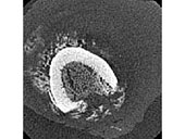
In the experiment involving the bone lengthening technique with the external fixation device, the internal structure of the bone was observed using the micro X-ray CT in 4 femur-cut rats in which serious drawbacks, such as detachment of the fixation device, severe purulence of the operated part due to infection, and marasmus due to indisposition, did not occur. The lateral image and the cross-sectional image obtained by using micro X-ray CT are shown in Figs. 5 and 6, respectively.
As shown in the lateral image, the bone was lengthened while maintaining the two cut surfaces parallel to each other. This indicated that the installation of the device and the bone lengthening were successfully completed. As shown in the cross-sectional images, the mesh structure that is typical of the woven bone that is formed at the beginning of the fracture healing process was observed. Therefore, the neogenetic bone tissue was considered to be favorably formed. |
4. Discussion
It was considered that detachment of flames in the experimental models (1)–(3) was due to the poor stability of flames against the bone. Since the purpose of this experiment was the formation of the neogenetic bone tissue with known load hysteresis, flames were affixed to the bone without using any bone cement. Therefore, carving of the bone in vivo with great accuracy was required; this was proved to be extremely difficult. Moreover, in order to widen the experimental region, the flames used were proportionally large; accidentally, the carved apertures became too large. In the experimental model (4), complete recovery of the cortical bone was not achieved. This could be due to adequate support of the rat’s body weight by the remaining bone tissues, leading to lesser load on the neogenetic bone tissue, and consequently a slower rate of tissue formation. Therefore, if the load on the neogenetic bone tissue is sufficiently large, the rate of tissue formation will be faster.
In the experimental model (5), the woven bone, which is a precursor of neogenetic bone tissue, was observed. Therefore, if the experimental duration is sufficiently long, neogenetic bone tissue formation will be obtained.
5. Conclusion
In this study, basic experiments were performed to investigate the formation of bone tissue with a known load hysteresis, which is essential for quantitative evaluation of dynamic remodeling of the bone tissue. Based on the results, it was clarified that neogenetic bone formation could be induced by applying a bone lengthening technique using an external fixation device. In order to improve the success rate of the surgical operations, improvement of experimental equipments and operational procedures, and development of a system in which the dynamic load imposed on the bone affects the neogenetic tissue formation are required.
|

| [1]
|
***** |
| [2]
|
Richards, M, Huibregtse, BA, Caplan,
AI, Goulet, JA, Goldstein, SA, Marrow-derived
Progenitor Cell Injections Enhance New
Bone Formation during Distraction, J.
Orthopaedic Research , Vol. 17, 1999,
pp. 900-908. |
|
|
|