
|
| Development of the Catheter Simulator |
|
|
1. Abstract
Recently, an interventional radiology as a minimally invasive treatment has been attracting considerable attention. The safety of the therapies mainly depends on the experience of the physicians in navigating the catheter into the blood vessels.
Computer-based simulators for surgical operations provide an effective training environment without danger to patients. This study aimed to develop a real-time simulation system (catheter simulator) in which a computerized virtual operative environment was constructed for the neuro-endovascular treatment, and a physician could be trained in the technique of catheter navigation by using the images of blood vessels of patients.
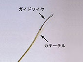
As the first step of this study, we constructed a physics-based model in which the bending and torsional deformations of a guidewire (a flexible wire positioned in a vessel for the purpose of directing the passage of a catheter, etc. (Fig. 1)) were considered. Moreover, we developed a novel computer-based training simulator for the neuro-endovascular treatment. |
|
|
| Fig. 1 A guidewire and a micro-catheter |
|
 |
|
|
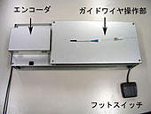
| Fig. 2 The input device |
|
|
|
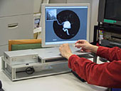
| Fig. 3 The catheter simulator |
|
2. The Catheter Simulator
The catheter simulator is a system (Fig. 3) consisting of an input device (Fig. 2) including the linear and rotational encoders, and a personal computer (PC). The simulator developed in this study allows the guidewire observed in the blood vessel displayed on the PC to be moved forward and deformed depending on the dynamic information computed using deformational and rotational magnitudes as detected by the input device; the movement and deformation of the guidewire can be displayed on the PC.
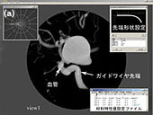
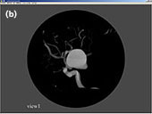
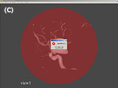
In our system, volume-based models constructed from MR/CT images of a patient's brain are used for the blood vessel modeling. The shape of the tip, initial location, and material characteristics of the guidewire are configured accordingly (Fig. 4 (a)). Subsequently, the guidewire is moved forward in the blood vessel using the input device or a mouse (Fig. 4 (b)). Any perforation on the vessel wall due to the guidewire can be judged based on the brightness value and a warning sign is displayed on the PC monitor (Fig. 4 (c)). |
|
|
| Fig. 4 Simulation of the guidewire |
3. A physics-based model for guidewire
(→ Download page (1))
The guide-wire is modeled by straight bar segments linked by virtual coil springs. In the physics-based model of the guidewire, only its quasi-static deformation was considered and the influence of blood flow was negligible. Moreover, only the bending and torsional deformations of the guidewire were considered as source of potential energy. The principle of stationary potential energy is used in order to identify the equilibrium solutions of the system. The vessel wall was expressed as an isosurface based on an arbitrary brightness value. Any contact between the guidewire and the vessel wall was judged based on the brightness value, and the normal vectors of the contact point were calculated from the gradient of the brightness values.
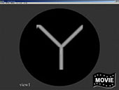
Figure 5 shows a computational example using a Y-shaped blood vessel model. It was confirmed that the guidewire was in contact with the vessel wall and moved forward along the vessel wall. |
|
|
|
Fig. 5 A computational example using a Y-shaped blood vessel model
(Click here to load the movie)
|
|
4. Conclusion
Since the catheter simulator does not use the FE mesh to express the vessels, it takes lesser time to input patient data obtained from MRI, etc., into the simulator. Furthermore, physicians can be trained by using this simulator. Thus, this simulator can contribute substantially to the development of appropriate treatment regimes for patients and the construction of an efficient and effective training environment for the physicians.
In the future, this simulator will be further improvised upon to enable its use for coupled analysis based on the deformation of the catheter and blood flow for constructing a virtual reality operative environment in which the input device can use the contact force between the guidewire and the vessel wall as feedback information.
|