
Three-dimensional internal structure microscopy (3D-ISM) (--> Hideo Yokota’s home page) enabled us to obtain full-color continuous cross-sectional images of a living body.
A biological model with color information is expected to be constructed by using 3D-ISM. In order to discriminate the different regions in the image, each tissue region must be split. However, the previous studies on region splitting in biological images were mostly performed for monochrome images, and the results obtained by these studies cannot be applied to the region splitting of a full-color biological image.
Region splitting in full-color biological images is still performed manually (--> Nobunori Kakusho’s home page); however, because of the enormous amount of data entailed, the automation of this process is urgently required.
In this study, a region splitting method suitable for full-color biological images was developed. |
|
|
|
|
The region splitting method includes a method that uses an edge in an original image, a region-based method, and a statistical method. In the method that uses an edge, the required edge does not always form a closed curve. Therefore, it is very difficult to discriminate the target region in biological tissues where different tissues lie close together.
In this study, a region splitting algorithm was developed, in which the region-based extraction method and the statistical method were combined. |
|
|
|
|
In this study, a method based on the region growing method was developed. In the region growing method, a region is split by successively integrating subregions into a region when a subregion of interest has the same characteristics as an adjoining subregion (or pixel).
In this study, the amount of characteristics is expressed by the HSV color coordinate system that is converted from the color information of the pixels. This is necessary because when the color of the biological tissue is expressed by the HSV color coordinate system, invariance is often observed in the same tissue. In addition, linear discriminant analysis was applied to discriminate between regions with the same characteristics. |
 |
|
|
|
|
In this experiment, mouse cross-sectional images and human eyeball cross-sectional images are used, which were obtained by using 3D-ISM.
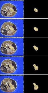
In the mouse cross-sectional images, the gastric region was split and region splitting was accomplished based on 100 continuous cross-sectional images (Fig. 1).
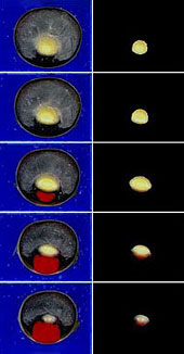
In the human eyeball cross-sectional images, region splitting of the lenticular region was accomplished (Fig. 2).
In this experiment, the extraction accuracy was greater than 90% throughout all the cross-sectional images; this result is higher than that obtained manually by people with anatomical knowledge (--> Nobunori Kakusho’s home page). (--> a list of related papers (6))
In the human eyeball cross-sectional images, a manually split region image was added once in every 20 cross-sectional images. However, this is a lesser work load compared to previous region splitting where a region in all cross-sectional images was manually split.
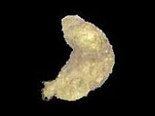
The abovementioned experimental results were visualized three-dimensionally by using the volume rendering method (Figs. 3 and 4).
|
|
|
| Fig. 1 The region splitting of the mouse gastric region (from 100 continuous cross-sectional images) |
Fig. 2 The region splitting of the lenticular region in the human eyeball (from 70 continuous cross-sectional images) |
|
|
|
| Fig. 3 The 3D model of the mouse gastric region |
Fig. 4 The 3D model of the lenticular region |
|
In the region splitting of full-color biological images, when the physical object is changed, the method must also be changed. In particular, the discriminating element in the algorithm and the amount of characteristics must be selected appropriately. Therefore, a database of the optimum method for various tissues obtained by 3D-ISM needed to be constructed.
In future, we will aim to construct an expert system on the region splitting of full-color biological images.
|