
|
| Determination of the Three-Dimensional Shape of a Human Eyeball with High Accuracy |
|
|
|
1. Method
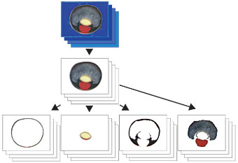
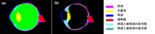
Using the three-dimensional internal structure microscopy (3D-ISM) developed by the Institute of Physical and Chemical Research (--> Hideo Yokota’s home page), a human eyeball was sliced at a 10 μm scale by a freeze-embedding method, and each sliced section was photographed using a high-definition camera with a resolution of 25μm (--> Sakiko Nakamura’s home page). Based on continuous cross-section images with full color image data obtained from the sliced sections of the eyeball, regions of each tissue (the whole eyeball, sclera, cornea, choroid + retina + ciliary body + iris, crystalline lens, anterior chamber + vitreous body, and optic nerve) in the eyeball were extracted by referring to anterior and posterior images and anatomical knowledge of these regions (Fig. 1). The image data on each tissue obtained from the region of extraction were converted into volume data, and continuous cross-section images parallel to the sliced sections were prepared. Subsequently, an incorrect image was amended by referring to its anterior and posterior images. The converted volume data on each tissue were visualized by a volume rendering/ray casting method (Voxel Viewer and Voxel Viewer 2; Toshiba Machine Co., Ltd., Japan).
|
 |
2. Results
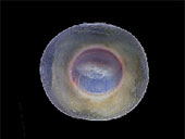
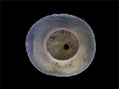
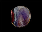
A stereoscopic image of each tissue could be observed by visualizing an arbitrary cross-section image with the axis between the top of the cornea and the center of the optic nerve endings (Fig. 2). Based on the transverse stereoscopic image of the sclera (Fig. 3), the position of the optic nerve could be determined. Since the position of the optic nerve in this image was shifted to the right side, this eyeball could be judged to be a right eyeball. Based on the stereoscopic cross-section image of the choroid + retina + ciliary body + iris (Fig. 4), the structure of the retina could be determined. Moreover, the shape of the ora serrata, which is the edge of the pars optica retinae, could be confirmed. Furthermore, other fine shapes in an eyeball could also be observed.
|
3. Conclusion
Based on the continuous cross-section images of a human eyeball that were obtained with high accuracy by 3D-ISM, the volume data on the whole eyeball, sclera, cornea, choroid + retina + ciliary body + iris, crystalline lens, anterior chamber + vitreous body, and optic nerve could be constructed, and these tissues could be observed stereoscopically. Since each tissue was extracted from the same images, the coordinate axes of all tissues were the same. Therefore, the position, size, and structure of each tissue could be compared. Moreover, since the cross-section image provided the internal information on an eyeball, a cross-section that was not actually sliced could be obtained, and each tissue could be observed. Since the region of each tissue was extracted in a manner that neighboring tissues were in contact with each other at the adjacent regions, no tissues overlapped. By adding pseudo-colors to the volume data constructed for each tissue, the position of each tissue could be distinguished clearly (--> Shinobu Hirata’s home page).
A geometric model for the simulation program could then be created using the volume data on each tissue, and an FEM mesh model could be constructed (Fig. 5).
|
|
Fig. 5 An image and a mesh model
(a) An image and (b) a mesh model |
|
|
|