
|
| Development of Three-Dimensional Internal Structure Microscopy (3D-ISM) and the Digitization of Biological Specimens |
|
|
Three-dimensional internal structure microscopy (3D-ISM) is a technique proposed by Higuchi et al. (--> a list of related papers (1)) that enable the internal structure of a specimen to be clarified without necessity of destructive sampling. The three-dimensional structure of a specimen can be observed with high accuracy and speed. The specimen is continuously sliced off and the remaining cross-section images are sequentially observed. Continuous images at a micron scale can thus be obtained (--> Hideo Yokota’s home page).
1. Macro-observation facility: digitization of an eyeball
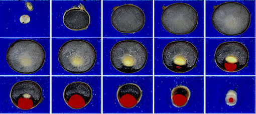
In this study, 3D-ISM was used to obtain biological data on shape, which are essential in order to create a simulation model for retinal detachment (--> Zhi-Gang Sun’s home page); a total of 3300 continuous cross-sectional images at a micron scale were obtained. Figure 1 shows 15 cross-sectional images obtained in this study.
The resolution was 10 μm along the thickness direction (Z-axis) and 25 μm along the plane direction (XY-axes); volume data with a resolution of 25 μm were obtained. These resolutions were sufficient to digitize a 100-μm thick retina. A stereoscopic image or an arbitrary cross-sectional image of the eyeball, the crystalline lens, and the cornea could be constructed by using the images obtained (--> Nobunori Kakusho’s home page). Based on these results, this facility is considered to be suitable for the digitization of the eyeball with high accuracy.
In addition, a shape model and a hexahedron finite element method (FEM) mesh model were created based on the data obtained (--> Shinobu Hirata’s home page).
2. Digitization of the blood circulatory system in a minipig
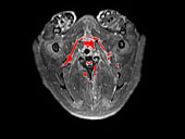
In order to construct a computer simulation model based on the shape data of the heart, the aortic arch, and the lungs in a minipig, the actual shape of the minipig was measured. After the blood was replaced with gelatine containing a fluorescence reagent and the specimen was sterilized, MRI (EXCELART MRT-2000/P2; Toshiba Medical System Corporation, Japan) was performed. Figure 2 shows the area vasculosa on the image obtained by the particular extraction method (a list of related papers (21)).
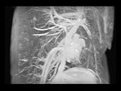
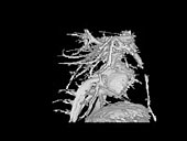
lthough the maximum intensity projection (MIP) image (Fig. 3) is only a tracing image of the entire volume data based on the visual point of the observer, the stereoscopic image (Fig. 4)—constructed by the ray-casting method based on the data obtainedclearly shows the three-dimensional structure of the heart and its surrounding blood vessels. Thus, continuous volume data could be obtained from the heart and its peripheral region by the digitization of the blood circulatory system.
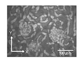
3. Micro-observation facility: the observation of capillary vessels in a small animal
An observation facility was developed in which a confocal laser scanning microscope (CLSM) was incorporated in its observational part. A CSU-10 confocal unit (Yokogawa Electric Corporation, Japan), which can obtain images in real time, was used for the confocal optics, and an ICCD camera (ICCD-300/DF; Hamamatsu Photonics K. K., Japan) was used for observation under low-intensity light conditions (--> Hideo Yokota’s home page).
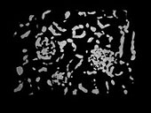
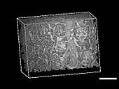
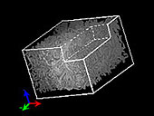
In order to observe a microscopic region of a biological specimen, casting resin was injected into a microvessel of a mouse kidney. The image of the microscopic three-dimensional structure obtained by the CLSM clearly shows the glomerulus and its surrounding blood vessels (Fig. 5). The resolution was very accurate—with XYand Z resolutions of 0.7 μm and 2 μm, respectivelyand 500 cross-sectional images were obtained within 5 min. Moreover, the microvascular system (Fig. 6) was extracted by the same method as that used for the minipig and the three-dimensional structure was visualized by the ray-casting method. Consequently, the three-dimensional connection of blood vessels and the arbitrary cross-sectional image could be observed (Figs. 7 and 8). Thus, a three-dimensional microscopic structure in a living body could be observed by 3D-ISM with CLSM.
This method can overcome a limitation of CLSM—the observation limit in the thickness direction—and, in comparison with the conventional scanning electron microscope (SEM) observations using casting resign injection, it has the advantage of clarifying the internal structure of biological specimens. Moreover, the volume data applicable to the simulation could be obtained by the extraction of microvessels (a list of related papers (30)).
|
|
|
| Fig. 5 |
|
|
|
| Fig. 6 |
|
|
 |
|